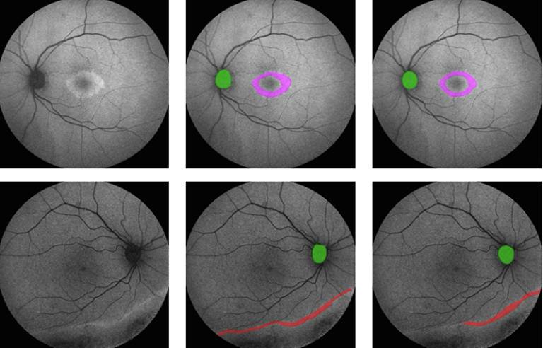
 Researchers at the UCL Institute of Ophthalmology and BRC have developed a deep learning model, ‘AIRDetect’ to process retinal imaging (fundus autofluorescence – FAF), a critical imaging tool for diagnosing and managing inherited retinal diseases. Interpretation of FAF imaging requires specialist knowledge, is prone to human-to-human variability, and is time-consuming. The new AI model provides a scalable, efficient, and rapid solution, which is also more reliable.
FAF imaging can detect retinal abnormalities at an early stage, key for timely and accurate diagnosis. This large-scale study successfully analysed a total of 45,749 retinal images from 3,606 Moorfields patients with a diverse range of inherited retinal conditions allowing for more data-driven comparisons. Previous research in this area has been limited to single conditions due to the large number of images to review. The study highlights the potential of AI-driven research to uncover patterns that were previously too time-consuming or too complex to analyse.
Further information
- View the full article on Ophthalmology Science
- Dr William A Woof
- Professor Michel Michaelides
- Professor Konstantinos Balaskas
- Associate Professor Nikolas Pontikos
- Image: Section from Figure 3 on published paper: Examples of manually and automatically segmented masks for the 5 features: vessels, disc, ring, hyper-autofluorescence (hyper-AF), and hypo-autofluorescence (hypo-AF). The vessel data set was separate from the rest of the data; hence, vessel visualization is separate from other features.