
Presenting on the recent CATION meeting was an excellent opportunity to connect with other researchers and clinicians, share my work, and gain insights that will certainly shape the next steps of my PhD journey. I spoke about my work called: “Assessing contrast sensitivity function in CRB1-retinopathies: Exploring child-friendly measures of visual function”. It aimed to explore contrast sensitivity function (CSF) across different spatial frequencies in patients with CRB1-retinopathies using the novel child friendly PopCSF test—an iPad-based, “gamified” assessment.
Mutations in the CRB1 gene are known to cause a spectrum of retinal dystrophies, including early onset severe retinal dystrophy/Leber congenital amaurosis (EOSRD/LCA), retinitis pigmentosa (RP), cone-rod dystrophy (CORD), and macular dystrophy (MD) leading to varying degrees of visual impairment. While there is currently no treatment for CRB1-retinopathies, research strategies towards treatment are progressing, making it crucial to establish reliable outcome metrics, especially for the paediatric population. In our prospective cross-sectional study, we evaluated 20 patients, including children, with confirmed biallelic CRB1 pathogenic variants at Moorfields Eye Hospital. We measured their best-corrected visual acuity (BCVA), contrast sensitivity using the Pelli-Robson chart, and CSF with the child friendly PopCSF test. The cohort included patients with EOSRD/LCA, CORD, and MD, allowing us to compare CSF across different CRB1-related phenotypes.
While there was no significant difference in the mean BCVA across the different phenotypes, we observed a significant difference in contrast sensitivity function. Pelli-Robson chart, the current gold standard measure for contrast sensitivity, and novel PopCSF test showed a moderate positive correlation (r=0.53, p=0.020). However, unlike the Pelli Robson chart, which only measures contrast sensitivity at a single spatial frequency, the PopCSF revealed clear losses in contrast sensitivity across both low and high frequencies, particularly in patients with EOSRD/LCA who exhibited the lowest CSF. This holds significant relevance since regulatory bodies are open to considering changes in contrast sensitivity across multiple frequencies for drug approvals. Therefore, CSF could emerge as a key metric for assessing visual function in CRB1-retinopathies.

.
B. A depiction of the contrast sensitivity function, shown with a sine-wave grating stimulus where its spatial frequency increases along the x-axis and contrast/amplitude decreases on the y-axis. The constructed area under the contrast sensitivity function (AUCSF) is depicted by the blue line, with contrast sensitivity plotted on the y-axis and spatial frequency (sf) on the x-axis (which should correspond with the point at which the depicted sine wave becomes difficult to detect against the background). The yellow circle shows the Gmax which is the peak contrast sensitivity and its spatial frequency (Fmax). β indicates the angle of decline of the CSF after Gmax (courtesy of Crossland MD et al. “Can children measure their own vision? A comparison of three new contrast sensitivity tests”)
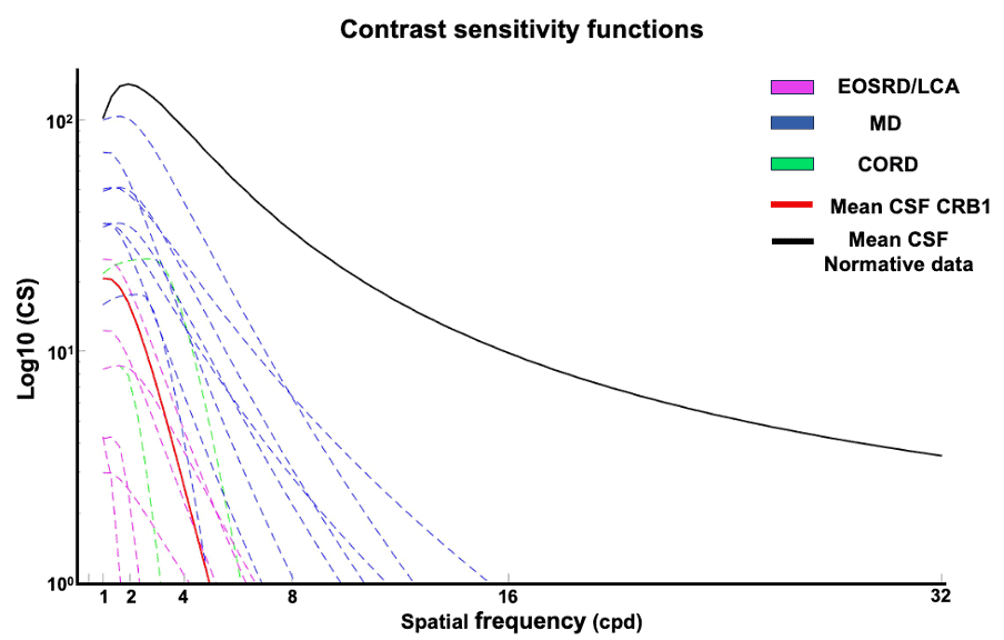
The CRB1 cohort had lower logCS at both low and high spatial frequencies compared to normative data, depicting an overall significantly lower CSF. Among the CRB1 phenotypes, patients with MD, indicated in blue, exhibited higher logCS at lower spatial frequencies compared to the other phenotypes, with the slope declining relatively consistently across different spatial frequencies thereafter. Contrastingly, patients with EOSRD/LCA, depicted in magenta, demonstrated the lowest CSF, by exhibiting the lowest logCS at lower spatial frequencies and a subsequent sharp decline at low spatial frequencies, indicating substantial losses in sensitivity to high frequencies. Similarly, the CORD group, represented in green, showed a sharp decline with increasing spatial frequency.
The discussions that followed my presentation were particularly enriching, especially on how these results could translate into clinical practice and their relevance to other retinal conditions. It was rewarding to see the level of interest and engagement from others working in this field, and I finished the meeting with a renewed sense of purpose and a few new ideas to incorporate into my research. I am excited to continue this research and to see how our findings might eventually contribute to better outcomes for patients with CRB1-retinopathies.